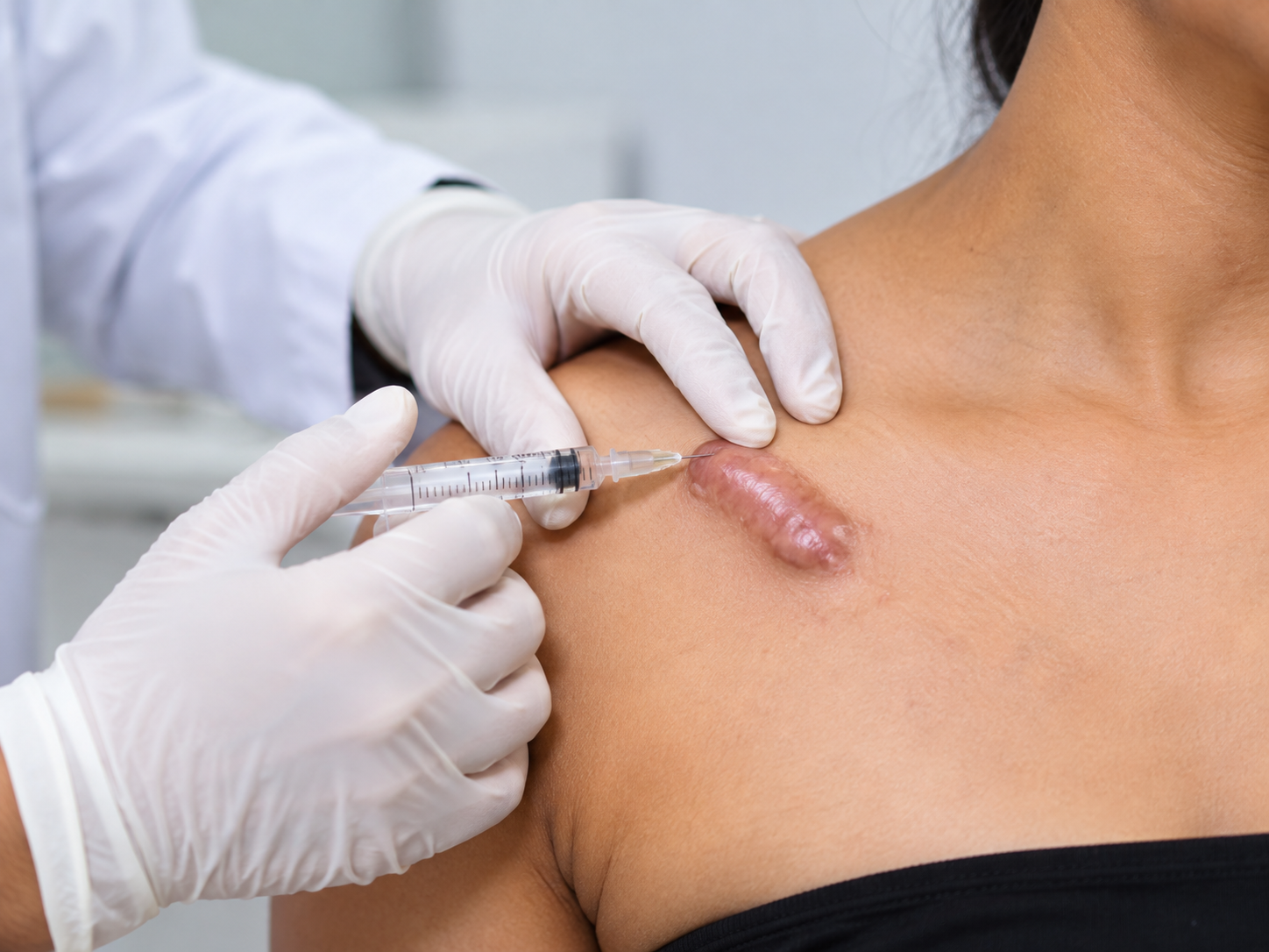
Keloid injection training for doctors should teach general practitioners how to assess suitable scars, understand intralesional triamcinolone concentrations, select the correct tissue plane, monitor adverse effects, and recognise when specialist referral is necessary. Published protocols commonly use 10–40 mg/mL triamcinolone at intervals of approximately four to six weeks, but the dose must be individualised and administered only by appropriately trained clinicians.
Keloids develop when scar tissue grows beyond the original wound boundaries. They may cause itching, pain, tenderness, cosmetic concerns and restricted movement.
Intralesional corticosteroid treatment can reduce scar thickness and symptoms in selected patients. However, incorrect technique or excessive steroid exposure may cause skin thinning, pigment changes and other complications.
Doctors therefore need practical training that combines procedural knowledge with patient selection, informed consent and complication management.
At Elegance Vidhyalay, training emphasises safe clinical assessment and supervised procedural education. Dr. Ashutosh Shah, M.S., M.Ch., D.N.B. (Plastic Surgery), a Plastic, Reconstructive and Cosmetic Surgeon with over 22 years of experience, contributes expertise in scar management, reconstructive principles and complication prevention.
General practitioners with appropriate training, professional authorisation and access to emergency support may be able to manage selected uncomplicated keloids.
However, not every raised scar is a keloid, and not every keloid is suitable for injection in a general outpatient setting.
A relatively straightforward case may involve:
Before recommending treatment, doctors should document the scar’s location, size, thickness, symptoms, and previous treatments.
Clinical photographs can help monitor changes over time.
Doctors should defer treatment or refer when they encounter diagnostic uncertainty, extensive disease, unusual growth, recurrent keloids or scars in anatomically challenging areas.
A lesion that bleeds unexpectedly, ulcerates, changes rapidly or has an unusual appearance requires further assessment before corticosteroid injection.
Doctors should also review allergies, active infections, diabetes control, medications and other relevant medical conditions.
A short training course alone does not establish independent competence or expand a doctor’s legally permitted scope of practice.
Intralesional triamcinolone acetonide is a commonly used corticosteroid for treating keloids.
It acts by suppressing inflammation and influencing collagen production within the scar.
The intralesional triamcinolone keloid dose depends on lesion thickness, treatment area, patient characteristics, and previous response.
Published guidance describes concentrations ranging from approximately 10 mg/mL to 40 mg/mL for selected hypertrophic scars and keloids.
Thicker lesions may require different treatment planning from thinner scars. However, doctors should not assume that the highest concentration will produce the best outcome.
Concentration describes the amount of medication in each millilitre of solution.
Total dose describes the amount of medication actually administered.
These measurements are different.
Doctors must record both concentration and injected volume so they can calculate and document total drug exposure.
A 2024 international expert consensus, the KECORT study, reported agreement on a preferred triamcinolone concentration of 40 mg/mL and a maximum of 80 mg per month in the specialist context it examined. This is not a universal GP dosing recommendation or an automatic maximum for every patient.
The responsible clinician must establish a patient-specific dose limit based on current evidence, product information and the approved treatment protocol.
| Keloid thickness | Published concentration reference | Volume guide | Typical interval | Stop or refer criteria |
|---|---|---|---|---|
| Small or relatively thin | Lower concentrations may be considered | Individualised by clinician | Approximately 4–6 weeks | Skin thinning, pigment change or diagnostic uncertainty |
| Moderate thickness | Commonly around 10 mg/mL in published scar protocols | Record planned and actual volume | Approximately 4–6 weeks | Poor response or increasing adverse effects |
| Thick, firm keloid | Specialist literature includes up to 40 mg/mL | Specialist-directed | Individualised | Extensive lesion, significant pain or high cumulative exposure |
| Large or recurrent keloid | Specialist assessment required | Not a routine GP protocol | Specialist-directed | Refer for combined treatment |
| Atypical lesion | Do not select steroid dose before diagnosis | Not applicable | Not applicable | Refer for diagnostic assessment |
Important: This table is an educational planning aid, not a prescribing chart. It deliberately does not assign a fixed injection volume to each scar category. The treating clinician must determine and document a safe total dose.
A central part of keloid injection training for doctors is understanding where the medication belongs.
Intralesional corticosteroid should enter the scar tissue itself rather than the surrounding normal skin or deeper subcutaneous fat.
Injecting into the subcutaneous tissue increases the risk of local tissue atrophy and visible depressions.
Keloids contain dense collagen tissue.
This firmness can make injection technically challenging and increase discomfort.
Doctors should learn to identify scar boundaries, understand tissue resistance, and recognise when a lesion is unsuitable for routine outpatient treatment.
Practical education should include:
The procedure should be taught through supervised clinical practice rather than attempted independently after reading a written technique description.
Doctors can explore Reconstructive surgery training to understand the broader principles of scar assessment, tissue healing and reconstructive management.
Doctors considering a dermatology procedures course in Valsad should evaluate whether the programme teaches patient safety and complication management alongside practical procedural skills.
An appropriate training programme should cover scar diagnosis, corticosteroid pharmacology, anatomical assessment, consent, dose documentation, injection-plane awareness, and postoperative review.
Trainees should also learn how to distinguish hypertrophic scars from keloids.
Hypertrophic scars generally remain within the boundaries of the original injury, while keloids extend beyond them.
This distinction matters because treatment response and recurrence patterns may differ.
A doctor should be able to:
Doctors interested in broader reconstructive education can review the Reconstructive surgery fellowship.
The Career options after MBBS without PG resource also provides information for medical graduates exploring further professional development.
Training eligibility must always be distinguished from independent procedural authorisation.
Intralesional steroid treatment can produce useful scar improvement, but adverse effects may occur.
Steroid-related atrophy causes thinning of the skin or underlying tissue.
It may appear as a depression near the treated scar.
Risk increases when corticosteroid reaches normal tissue or subcutaneous fat, or when cumulative exposure is excessive.
Doctors should stop and reassess treatment when atrophy develops.
Patients may develop hypopigmentation or other changes in skin colour around the injection site.
These changes can be especially noticeable in darker skin tones.
Doctors must discuss this possibility during consent and assess pigment changes at follow-up.
Small superficial blood vessels may become more visible after repeated steroid exposure.
This may indicate local skin changes requiring treatment reassessment.
Dense keloid tissue can make injections uncomfortable(keloid injection training for doctors).
Pain management should follow an appropriate clinical protocol and account for allergies, local anaesthetic exposure and patient tolerance.
Doctors should not assume that combining medications automatically eliminates discomfort.
Although uncommon, infection, ulceration and other local reactions may occur.
Treatment should be postponed when the injection site has an active infection.
Unexpected worsening, severe pain, progressive ulceration, or concerning skin changes require prompt clinical evaluation.
Some keloids require more than steroid injections.
Specialist referral is particularly important for:
Keloids can recur after excision.
Surgical treatment may therefore require an individualised postoperative plan involving scar prevention and additional therapies.
Depending on the lesion, specialists may consider corticosteroid injections, silicone therapy, pressure treatment, selected laser procedures, 5-fluorouracil or other approaches.
The International Advisory Panel on Scar Management supports individualised, multimodal treatment for difficult scars.
These treatments differ in their risks and required expertise.
A GP should not begin specialist combination therapy without the necessary qualifications, training and clinical support.
Keloid recurrence prevention requires realistic counselling and long-term follow-up.
Doctors should explain that successful flattening does not guarantee permanent resolution.
Patients may require further assessment if the scar becomes raised, painful or itchy again.
A silicone sheet after keloid injection may form part of a broader scar management plan when the skin is intact, and the treating clinician considers it appropriate.
Silicone gels and sheets are widely used non-invasive options for scar prevention and treatment.
They should not be placed over an open or infected wound without appropriate medical guidance.
Doctors can review the experience and qualifications of Elegance Vidhyalay’s Faculty when evaluating available training opportunities.
The number of injections varies according to scar thickness, location, and response. Some patients require several sessions spaced approximately four to six weeks apart. Doctors should reassess the scar after each session rather than commit to a fixed number of injections regardless of response.
Some published protocols describe dilution with a local anaesthetic, but compatibility, concentration, and total drug exposure must be considered. Doctors should follow product-specific guidance and an approved clinical protocol rather than mix medications routinely without appropriate assessment.
Intralesional corticosteroids can cause local skin or fat atrophy. The risk increases when medication reaches surrounding normal tissue or deeper subcutaneous fat. Doctors should assess any developing depression or thinning and reconsider further treatment.
5-fluorouracil is used in selected specialist scar-management protocols, sometimes alongside corticosteroids. However, it is a cytotoxic medication requiring appropriate expertise, precautions, and monitoring. Doctors exploring a dermatology procedures course in valsad should learn when specialist referral is preferable to attempting advanced combination treatment.
Silicone sheets or gels may be introduced when the skin surface is intact, and the treatment plan supports their use. They can complement other scar treatments, but the schedule depends on wound healing, skin tolerance and clinical assessment.
The timing depends on the surgical procedure, wound healing, and recurrence-prevention plan. Some specialist protocols include perioperative or postoperative corticosteroid treatment, but there is no universal schedule suitable for every patient. The operating surgeon should determine the appropriate timing.
Keloid injection training for doctors should begin with accurate diagnosis, patient selection, and knowledge of steroid-related risks.
Intralesional triamcinolone concentrations commonly discussed in published guidance range from 10 to 40 mg/mL for selected scars, but concentration and total dose must be assessed separately.
The injection must remain within the appropriate scar tissue plane to reduce the risk of skin and subcutaneous fat atrophy.
Doctors should monitor pain, pigment changes, telangiectasia, thinning and treatment response at follow-up.
Large, recurrent, anatomically challenging or atypical keloids require specialist assessment.
Silicone therapy and other recurrence-prevention measures may complement treatment when clinically appropriate.
At Elegance Vidhyalay, structured training should help doctors understand both procedural principles and the limits of their clinical competence.