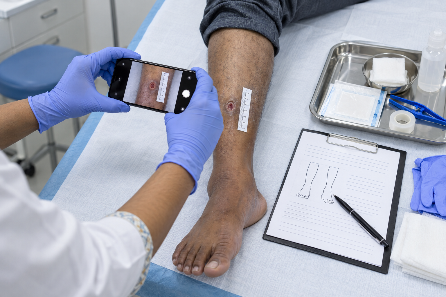
A consistent clinical photography protocol for wounds helps doctors track healing, compare changes between visits, communicate findings, and maintain a reliable clinical record. Photograph every wound at the same distance, angle, and lighting, with a disposable scale marker and anatomical landmark visible. At every visit, record the wound’s length, width, and depth in millimetres and store photographs within the clinical record rather than on a personal phone.
Good wound documentation combines written clinical findings, measurements, photographs, treatment details, and follow-up plans. Photography should support the written record, not replace it.
Each dressing or wound-care visit should create a clear record of the patient’s current condition and any changes since the previous assessment.
A consistent template makes comparison easier and reduces missing information.
| Field | What to Record |
|---|---|
| Date and time | Date and time of assessment |
| Wound location | Exact anatomical site |
| Wound type | Clinical description/classification where appropriate |
| Length | Measurement in millimetres |
| Width | Measurement in millimetres |
| Depth | Measurement in millimetres |
| Wound bed | Relevant wound-bed findings |
| Wound edges | Condition of margins/edges |
| Surrounding skin | Relevant periwound findings |
| Exudate | Amount and relevant characteristics |
| Odour | Presence or absence where clinically relevant |
| Infection findings | Relevant local/systemic findings |
| Pain | Patient-reported pain where relevant |
| Vascular/neurological findings | When clinically indicated |
| Photograph | Standardised clinical image |
| Treatment | Cleansing, dressing or procedure performed |
| Offloading | Method used or advised where relevant |
| Advice | Patient instructions |
| Follow-up | Review date and escalation instructions |
| Clinician | Name/identification of person completing record |
The clinic should use the same structure at each visit wherever practical.
Doctors developing documentation systems can also review Practice management for doctors.
Measurement becomes useful only when the team uses a consistent technique.
The supplied protocol requires recording:
Length × width × depth in millimetres.
The clinician should clearly identify which dimension represents length and width and use the same measurement convention at subsequent visits.
Do not rely only on photographs to estimate wound dimensions.
Record measurements directly in the clinical notes.
Suppose different staff members measure the same wound using different orientations or techniques. Apparent changes may reflect measurement variation rather than actual healing or deterioration.
A clinic should therefore establish one measurement method and train everyone involved in wound documentation to follow it.
Clinical photographs become easier to compare when every image follows the same protocol.
The objective is repeatability.
| Element | Rule | Why It Matters |
|---|---|---|
| Distance | Use the same practical camera-to-wound distance at follow-up visits | Reduces apparent size differences caused by camera position |
| Angle | Photograph from the same angle each time | Makes serial images easier to compare |
| Lighting | Keep lighting consistent and minimise distracting shadows or glare | Helps preserve visual consistency |
| Scale placement | Place a disposable scale marker beside the wound | Provides a size reference |
| Anatomical marker | Include a recognisable anatomical landmark when appropriate | Helps confirm wound location and orientation |
| Background | Use a clean, consistent and non-distracting background | Keeps attention on the clinical subject |
| File naming | Follow the clinic’s standard patient/image identification system | Reduces filing errors |
| Storage location | Store the image within the authorised clinical-record system | Keeps photographs connected to the patient’s record |
Moving the camera closer makes the wound appear larger. Moving farther away makes it appear smaller.
Therefore, avoid taking one photograph as an extreme close-up and another from considerably farther away.
The clinic can develop a standard practical distance for particular types of wound photographs.
Consistency matters more than creating a visually dramatic image.
Photographing a wound directly during one visit and obliquely during the next can distort comparisons.
Use a repeatable orientation whenever possible.
For difficult anatomical areas, document the position used so staff can reproduce it during follow-up.
Lighting can significantly change how tissue appears in a photograph.
Avoid:
Use neutral, consistent clinical lighting whenever possible.
The objective is documentation, not photographic enhancement.
The supplied protocol recommends using a disposable scale marker with wound photographs.
Place the scale close to the wound without obscuring important clinical details.
The scale provides a visual reference, but it does not replace documented wound measurements separately.
A useful sequence is:
Measure → record dimensions → position scale → photograph → store image.
An isolated close-up may make it difficult to identify exactly where the wound is located.
Including an anatomical landmark can provide useful context.
In some cases, the clinic may take:
The same approach should then be repeated during subsequent visits where appropriate.
Patients should understand why the clinic wants to take photographs and how it intends to use them.
Clinical documentation and public use are not the same purpose.
Consent documentation should clearly distinguish between images taken for the clinical record and any proposed secondary use.
The record should document the relevant consent process according to the clinic’s policy and applicable requirements.
Do not assume that permission to take a clinical photograph automatically authorises its use for teaching, websites, social media, advertising, or other promotional purposes.
Those uses create different considerations.
If a clinic wants to use photographs beyond direct clinical documentation, it should follow an appropriate separate consent process and applicable privacy and professional requirements.
Clinical care should not depend on a patient agreeing to promotional use of their images.
The supplied protocol specifically recommends storing wound images in the clinical record rather than on a phone.
A personal camera roll creates obvious organisational and privacy problems.
The preferred workflow is:
Take authorised clinical photograph → transfer/store it in the approved clinical system → verify correct patient record → follow the clinic’s device-handling policy.
Staff should not maintain uncontrolled collections of patient wound photographs on personal devices.
Clinical images form part of the patient’s documentation when the clinic uses them for clinical care.
The storage process should therefore protect:
The clinic should define who can access photographs and how staff add images to the patient record.
A backup process should protect authorised records against accidental loss while maintaining appropriate access controls.
Doctors and clinical staff interested in structured diabetic foot care can review the Diabetic foot technician course.
Random filenames make image management difficult.
A clinic should establish a standard naming convention within its authorised system while avoiding unnecessary exposure of patient information.
The naming system should help staff connect the image with:
The exact format should follow the clinic’s record-management system and privacy requirements.
The supplied blog plan does not specify a fixed retention period.
Clinical photographs that form part of the medical record should follow the applicable record-retention requirements for the practice and jurisdiction.
Clinics should therefore verify current legal, professional, institutional, and record-specific requirements instead of applying an arbitrary number of years.
The clinic’s retention policy should also address backup copies and authorised storage locations.
Good documentation creates a chronological account of clinical care.
If questions arise later, the record can show:
What the wound looked like → what its measurements were → what treatment the clinician provided → what advice the patient received → how the wound changed → what decisions the clinician made.
Photographs become particularly useful when they correspond with dated written notes and measurements.
However, images without context can create uncertainty.
For example, a photograph alone may not explain:
That is why photography should complement the clinical note.
Several simple mistakes can reduce the usefulness of serial wound photographs.
Changing distance: Staff photographs the wound from different distances at every visit.
Changing angle: Images show the wound from inconsistent perspectives.
No scale marker: The photograph provides no reliable visual size reference.
Poor lighting: Shadows or glare obscure relevant details.
No anatomical context: A close-up makes the wound location difficult to identify.
Personal-phone storage: Staff leaves clinical images outside the authorised patient record.
Unclear consent: The record does not clearly document the purpose of photography.
Using images for another purpose without appropriate permission: A clinical image later appears in teaching or promotional material without a separate appropriate process.
A written protocol can reduce these inconsistencies.
A practical workflow can help staff repeat the same process:
1. Review consent and patient identification.
Confirm that the correct patient and wound are being documented.
2. Assess and measure the wound.
Record length, width, and depth in millimetres.
3. Prepare the photograph.
Use consistent positioning, lighting, background, and camera distance.
4. Position the disposable scale marker.
Keep it visible without covering the wound.
5. Include anatomical context.
Include an appropriate landmark or take an additional contextual photograph where useful.
6. Take the photograph.
Avoid filters and unnecessary enhancement.
7. Store it correctly.
Place the image in the authorised clinical record rather than leaving it on a personal phone.
8. Complete the written note.
Record findings, treatment, dressing, advice, and follow-up.
A protocol works only when everyone follows it consistently.
Doctors, nurses, technicians, and other staff involved in wound care should understand the clinic’s measurement, photography, consent, and storage processes.
At Elegance Vidhyalay, training programmes can support doctors and healthcare professionals in developing structured clinical and practice-management skills.
Training associated with Dr. Ashutosh Shah, Plastic and Reconstructive Surgeon with 22+ years of clinical experience, also emphasises systematic clinical assessment and documentation alongside practical wound-care skills.
Doctors can explore available Programmes for doctors and the Institute image gallery.
The appropriate consent process depends on the purpose and applicable requirements. Clinics should clearly document consent for clinical photography and distinguish clinical-record use from teaching, marketing, or other secondary uses.
Use a consistent measurement method at every visit and record the wound’s length, width, and depth in millimetres. Apply the same measurement convention during follow-up assessments.
The supplied protocol recommends storing images in the authorised clinical record rather than keeping wound photographs on a personal phone.
Yes, the supplied protocol recommends a disposable scale marker. Place it beside the wound without covering relevant clinical features, while also recording measurements separately.
Follow the applicable record-retention requirements for the clinic and jurisdiction. Because requirements vary, verify current professional, legal, and institutional rules rather than assuming a fixed period.
Clinical-record consent should not automatically be treated as permission for teaching or marketing. Follow an appropriate separate consent process and applicable privacy and professional requirements for secondary use.
A reliable clinical photography protocol for wounds depends on consistency.
At every visit, record the wound’s clinical findings and length, width, and depth in millimetres. Photograph it using consistent distance, angle, lighting, scale placement, anatomical context, and background.
Most importantly, keep photographs within the authorised clinical record, maintain appropriate consent and access controls, and use a separate process when considering images for teaching or marketing.
When the whole team follows the same protocol, serial photographs become much more useful for monitoring wound progression and supporting complete clinical documentation.